|
|
|
| Prosthetic Rehabilitation For A Case Of Papillon-lefevre Syndrome |
Sujana Varri 1 , Ram Sunil Chukka 2 , Anupama Tadepalli 3 , Ravi Kiran 4
1 Reader,Dept Of Conservative Dentistry And Endodontics - Sibar Institute Of Dental Sciences, Guntur,A.P, India.
2 Professor, Dept Of Conservative Dentistry And Endodontics - Sibar Institute Of Dental Sciences, Guntur, A.P,India.
3 Reader, Dept Of Periodontics - Srm College Of Dental Sciences, Chennai, India.
4 Professor, Dept Of Prosthodontics - Dr Sudha Nageswararao Institute Of Dental Sciences,A.P,India.
|
| Address For Correspondence |
Dr. V.Sujana, D.No: 21-10-37/3,
Srinagar Colony II Lane,
Satyannarayana Puram, Vijayawada -520011.
Email : drsujanavarri@yahoo.co.in |
| Abstract |
| Papillon-lefevre syndrome is a rare syndrome of autosomal recessive inheritance characterized by palmo-plantar hyperkeratosis and a precocious progressive periodontal disease that results in premature loss of primary and permanent dentition. Here, is a case report of prosthodontic rehabilitation of a 15-year-old female with Papillon-lefevre syndrome. |
|
| Keywords |
| Cast partial denture, Hyperkeratosis. |
|
| Full Text |
Introduction
Papillon-lefevre syndrome was first described by Papillon & Lefevre in 1924.[1] The clinical manifestations include palmo-plantar hyperkeratosis and rapidly progressing periodontitis which results in premature exfoliation of primary and permanent dentitions. Gorlin et al suggested that calcification of duramater is the 3rd component of the syndrome. Prevalence of Papillon-lefevre syndrome is 1-4 per million individuals with no sex predilection and racial predominance. A greater frequency of occurrence is noticed in consanguineous offspring.
The eruption of primary teeth occurs at the normal age in a normal sequence with normal form and structure. Following eruption of primary teeth, severe gingival inflammation and a generalized aggressive periodontitis occur resulting in premature loss of primary teeth. Gingiva resumes its normalcy after exfoliation of the primary teeth. Permanent dentition is also followed by same events and if not intervened most of the permanent teeth are lost by 15-17 years of age. Severe resorption of alveolar bone gives the teeth a floating in air appearance on radiographs. Plamo-planar hyperkeratosis varies from psoriasiform scaly skin to hyperkeratosis and this can also affect other areas such as the elbows and knees.
Case Report
A 15-year-old female patient came to the dental office with a chief complaint of mobility of upper and lower anterior teeth and pain while taking food. Family history revealed consanguineous marriage of their parents. She is the second of the three siblings born after a full term normal delivery and none of her siblings showed similar findings.
She is thin built with a steady gait with normal physical and mental development. Dermatological examination revealed hyperkeratotic, well demarcated asymptomatic plaques extending on to the dorsal surfaces of hands and feet. She also gave a repeated history of skin infections.
Dental history revealed early loss of primary teeth. Intraoral examination revealed grade II mobility of 21, 31, 32 & 41. Deep pockets i.r.t 16, 26, 36 & 46. Panoramic view showed generalized loss of alveolar bone with complete loss of bone support giving a floating in air appearance.
Blood investigations and liver function tests showed values within normal range. Based on these findings the condition was diagnosed as Papillon-lefevre syndrome.
Treatment And Management
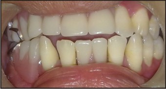
Since the patient is a teenage girl in order to satisfy the esthetic and functional needs extraction of the mobile teeth, periodontal therapy combined with prophylactic antibiotics of remaining teeth and replacement of missing teeth by using upper and lower cast partial dentures were planned.(Figure 1, 2) Course of antibiotics helps to control active periodontitis in an effort to preserve the teeth a and to prevent bacteremia and subsequent pyogenic liver abscess The patient was referred to a dermatologist for the management of skin lesions.
 | Pre Operative View
 |
 | Post Operative View
|
Discussion
Papillon-lefevre syndrome is a rare disorder of keratinisation affecting children between ages of 1-5 years. The reported incidence is 1-4 per million. The exact etiology and pathogenesis is unknown but three main factors are responsible for initiation and progression of Papillon-lefevre syndrome.[1]
Genetically loss of function, mutations of the lysosomal protease cathepsin C[2],[3],[4],[5],[6]. gene are associated with Papillon-lefevre syndrome and related conditions. The cathepsin C gene is expressed in the epithelial regions and in various immune cells including polymorphonuclear leucocytes, macrophages and their precursors. This gives better understanding of the signs and symptoms associated with Papillon-lefevre syndrome.[1]
Immunologically alteration of the host defense because of decreased function of PMNLs or monocytes.
Microbially gram negative microbial polysaccharides are recognized as the primary factors in the etiology of periodontitis in Papillon-lefevre syndrome. Actinobacillus actinomycetemcommitans constituted more than 50% of colony forming units.[1]
Plamo-planar keratosis starts within 4 years of life with sharply demarcated erythematous keratotic plaques involving palms and soles, sometimes extending on to the dorsal surfaces of hands and feet. The cutaneous lesions have a tendency to worsen in winter.[7],[8],[9]. Oral manifestations of Papillon-lefevre syndrome appear almost simultaneously with the onset of palmo-plantar hyperkeratosis. The primary teeth erupt at the expected age and in normal sequence.
Usually, the teeth are of normal form and structure. Rapidly progressing periodontits ensues, after the eruption of the primary dentition, manifested by markedly reddened, inflamed and swollen gingival associated with extensive bone resorption and deep periodontal pockets from which pus exudes in response to slight pressure. Chewing is very painful because of the mobility of teeth. Fetid mouth odour and regional lymphadenopathy are observed commonly.[10]. Increase susceptibility to infections was reported in 20 per cent patients due to dysfunction of leucocytes and neutrophils.[11]. Pyogenic liver abscess is increasingly recognized as a complication PLS associated with impairment of immune system.[12].
The dental features of Papillon-lefevre syndrome are the looseness, hyper mobility, drifting, migration and exfoliation of teeth without signs of root resorption. Primary teeth are exfoliated or extracted and child becomes completely edentulous by the age of 4-5 years with gingival returning to normal healthy state. Same cycle begins with the eruption of permanent teeth and by the age of 13-15 years if not intervened all permanent teeth are lost. Radiographic examination reveals severe loss of alveolar bone and teeth appear to be “floating in air”[1].
Differential diagnosis includes chediak-higashi syndrome, juvenile periodontitis and Haim – Munk syndrome. Features associated with chediak-higashi syndrome are absent in this case. Skin lesions are present by which only juvenile periodontitis can be ruled out. In haim – munk syndrome apart from features seen in PLS archanodactyly, acroosteolysis, atophic changes of nails and radiographic deformity of fingers can be seen.[5].
The Papillon-lefevre syndrome can adversely affect growing children psychologically, socially and aesthetically. A multi-disciplinary approach may improve the prognosis and quality of life of these children. Thus, oral rehabilitation in such patients is a must. Prosthetic replacement in Papillon-lefevre syndrome is age specific, specialty job involving initial replacement with complete or partial dentures with future consideration for implant supported prosthesis. In this case we have selected upper and lower cast partial dentures for prosthetic replacement because she is still young and loss of any tooth can be added to the framework in future. After the completion of growth we have recommended her for implant supported dentures.
Conclusion
Early recognition and a multi-disciplinary approach helps in improving the prognosis of patients with Papillon-lefevre syndrome. Prosthetic rehabilitation provides a psychological boost up to the patient and parents by restoring not only the esthetic appearance but also the function.
References
1. Parmanand J. Dhanrajani. Papillon-Lefevre syndrome: clinical presentation and a brief review. Oral Surg Oral Med Oral Pathol Oral Radiol Endod 2009;108: 1-7.
2. Thomas C Hart, P Suzanne Hart, Donald W Bowden. Mutations of the cathepsin C gene are responsible for Papillon-Lefèvre syndrome. J Med Genet 1999;36:881–887.
3. Mazen Kurban a Muhammad Wajid a Yutaka Shimomura. Evidence for a Founder Mutation in the Cathepsin C Gene in Three Families with Papillon-Lefèvre Syndrome. Dermatology 2009;219:289–294.
4. Caroline Lefevre, Claudine Blanchet-Bardon. Novel Point Mutations, Deletions, and Polymorphisms in the Cathepsin C Gene in Nine Families from Europe and North Africa with Papillon - LefeÁvre Syndrome. J Invest Dermatol 117:1657 - 1661, 2001.
5. P S Hart, Y Zhang, E Firatli, C Uygur. Identification of cathepsin C mutations in ethnically diverse Papillon-Lefèvre syndrome patients. J Med Genet 2000;37:927–932.
6. Aoi Nakano, Kazuo Nomura, Hajime Nakano. Papillon - Lefevre Syndrome: Mutations and Polymorphisms in the Cathepsin C Gene. J Invest Dermatol 116: 2001, 339 – 343.
7. Nagaveni N.B, Suma .R, Shashikiran N.D, Subbareddy V.V. papillon lefevre syndrome: report of two cases in the same family. J Indian Soc Pedod Prevent Dent June 2008; 78 – 81.
8. Ashwani P, Swapna K, Sailaja Rani M, B.S.N Reddy. Papillon – lefevre syndrome with pseudoainhum. Indian Dermatology online Journal, Dec 2010; (1), 33 – 35.
9. Aldevina C. de Freitas, Sada Assed. Aggressive periodontitis associated with Papillon- Lefevre syndrome: Report of a 14-year follow-up. Spec Care Dentist, 2007, 27(3): 95- 100.
10. Subramaniam.P, Mathew.S. Papillon –Lefevre syndrome: A case report. J Indian Soc Pedod Prevent Dent, Dec 2008: 171 – 174.
11. Jain V., Gupta K. Prosthodontic rehabilitation in Papillon –Lefevre syndrome: A case report. J Indian Soc Pedod Prevent Dent, June 2008: 95 – 98.
12. SS Dhanawade, SD Shah and GM Kakade. Papillon- lefevre syndrome with liver abscess. Indian pediatrics 46 Aug 17, 2009, 723 – 725.
|
|
|
|
|
|
|